that is rich in GC base pairs. More recently, it has
been suggested that the R-bands are regions of the
chromosome that are stretched during routine
chromosome preparation and that G-bands are
unstretched, and therefore DNA-rich, regions of the
chromosome; the banding pattern would then arise
from “a fixed hierarchy of the stretchibility of
chromosomes” (Hliscs
et al.
1997).
The individual chromosomes are identified by
size, position of the centromere, and the G-banding
pattern. The chromosomes are then arranged into a
karyotype wherein the chromosome pairs are set out
in numerical sequence. The sex chromosomes may
be paired separately or grouped with the chromo-
some pairs of similar size: the X chromosome with
chromosomes 6 to 12 (group C) and the Y chromo-
some with chromosomes 21 and 22 (group G).
Abnormalities of chromosome number are apparent
by a simple count. If there is an extra chromosome,
it is identified by the presence of a threesome rather
than a pair of one of the chromosomes. The XO
monosomy is identified by the presence of a single
sex chromosome, an X chromosome. Structural
chromosome abnormalities are detected as chromo-
somes which do not show any of the normal
G-banding patterns for a chromosome of their size
and centromere location. If the structural
abnormality involves one chromosome, the karyo-
type will show one abnormal chromosome and one
unpaired chromosome. By comparing the G-banding
pattern of the abnormal chromosome to the normal
pattern of the involved chromosome, the nature of
the abnormality can be characterized. For example,
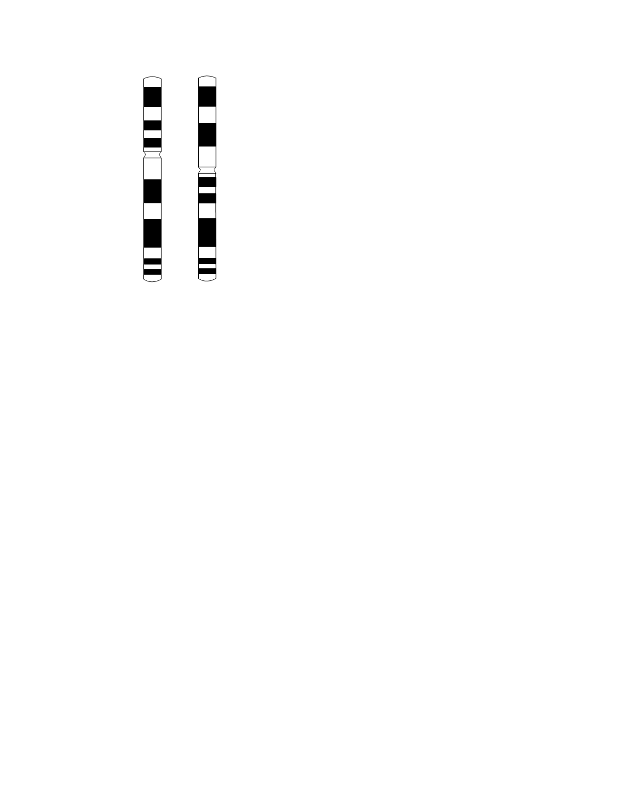
Figure 10.1 shows the G-banding pattern in the case
of an inversion of chromosome 7. It is clear that the
aberrant portion of the abnormal chromosome is
between band 15 of the short arm and band 22 of the
long arm. It is also clear that the abnormal portion
has a G-banding pattern that corresponds to an inver-
sion of the normal portion. If the structural abnor-
mality involves two chromosomes, the karyotype
will show two abnormal chromosomes and either
two unpaired chromosomes or a missing chromo-
some pair. Here also the abnormality can be charac-
terized by comparison with the normal G-banding
pattern of the involved chromosomes.
In fluorescence
in situ
hybridization, chromo-
some identification is accomplished by using single-
strand nucleotide probes that hybridize to chromo-
some-specific DNA sequences. The probes are
labeled with a fluorochrome (a fluorescent chemi-
cal). Because the fluorescence signal does not
require that the chromosomes be condensed in order
to be seen, this method can be applied to interphase
nuclei of nondividing cells. It can also be used as an
alternative technique for the analysis of metaphase
chromosome preparations.
The probes used for fluorescence
in situ
hybridi-
zation are of three types: chromosome-specific paint-
ing probes, centromeric repeat probes, and
locus-specific probes. Chromosome-specific paint-
ing probes hybridize to many sites along the length
of a single chromosome, thereby “painting” the
chromosome. To paint each of the 22 autosomes
and each of the sex chromosomes uniquely so that a
complete karyotype can be analyzed, 24 unique
colors are needed. This could be accomplished by
using 24 different fluorochromes but that number
exceeds the number of fluorochromes available.
Instead, by labeling each probe with a more than one
fluorochrome in a unique combination, 24 distinct
signals can be generated with many fewer fluoro-
chromes—only 5 are needed. Karyotype analysis
using this approach is called multiplex fluorescence
in situ
hybridization or spectral karyotyping (SKY).
For a number of full-color examples of the applica-
tion of this approach, the reader is directed to the
article by Speicher
et al.
(1996). The advantages of
Genetic Disease
10-2
21
22
31
32
12
13
14
15
21
22
33
34
35
36
p
q
normal
inversion
Figure 10.1
The G-banding pattern for chromosome 7 at a
resolution of 450 bands per haploid set. A normal chromo-
some 7 is shown on the left. The band numbering schemes
for the short (p) and long (q) arms are indicated. A chromo-
some 7 with a pericentric inversion is shown on the right.


