some defined portion of the fragment. The length of
a DNA fragment will be altered if a deletion, inser-
tion, or expansion of trinucleotide (triplet) repeat
sequences is present within the fragment. If the
alteration in fragment length is larger than 50 to 100
base pairs, it can be detected as a change from
normal in the electrophoretic location of the
fragment.
A change in the length of a DNA fragment can
also be caused by a nucleotide substitution that
creates or destroys a cleavage site for a restriction
endonuclease. For instance, the mutation that pro-
duces sickle hemoglobin (6 Glu
→
Val) fortuitously
alters the sequence at a site for the restriction
enzyme
Mst
II. Following incubation with
Mst
II and
using a probe to the 5’ flanking DNA of the
β
-globin
gene, normal DNA generates a 1.15-kb fragment
due to cleavage at codon 6. In contrast, sickle DNA
generates a 1.35-kb fragment as the next
Mst
II
restriction site is 200 base pairs downstream from
codon 6 (Figure 10.2). Unfortunately, most nucleo-
tide substitutions do not alter a restriction endonucle-
ase cleavage site. Southern blot hybridization can
still be used as a diagnostic approach for such
mutations by performing the analysis on DNA that
has been amplified by PCR using a primer that intro-
duces an allele-specific restriction enzyme cleavage
site (as discussed above).
Southern blot hybridization is also frequently
used to diagnose genetic disease when the gene
responsible for the disease has not been identified or
when the site and character of the mutation causing
the disease is unknown even though the gene
involved has been identified. In these circumstances
it is often possible to identify a polymorphic DNA
sequence that, while not itself being the site of the
mutation, is in close linkage with the mutation.
Hence the polymorphism and the mutation are, for
the most part, co-inherited and the presence of the
polymorphism serves as a marker of the mutation.
Polymorphisms that arise from nucleotide variability
at the site of a potential restriction endonuclease
cleavage site, called restriction fragment length
polymorphisms (RFLPs), are particularly useful.
For example, a mutation may be linked to DNA that
has a polymorphism characterized by the presence of
a novel endonuclease restriction cleavage site (as in
Figure 10.3). Following digestion with the appro-
priate endonuclease restriction and Southern blot
hybridization with a probe to DNA near the novel
cleavage site, the polymorphism will show a shorter
than normal DNA fragment. Conversely, if the
mutation is linked to a polymorphism that lacks a
endonuclease restriction cleavage site that is usually
present, Southern blot hybridization will reveal a
DNA fragment that is longer than normal. In either
case, the presence of the polymorphism as demon-
strated by Southern blot hybridization can be used as
a diagnostic marker for the linked mutation.
However, because genetic linkage is never absolute,
the diagnostic performance of polymorphisms is not
as good as that of direct molecular studies of the
mutation. Additionally, polymorphisms can only be
Genetic Disease
10-6
AA
AS
SS
Genotype
1.35
1.15
kb
Figure 10.2
Schematic depiction of the molecular diagno-
sis of the sickle hemoglobin mutation using Southern blot
analysis. The restriction endonuclease is
Mst
II and the
labeled oligonucleotide probe is complementary to a portion
of the 5’ flanking DNA of the
β
-globin gene.
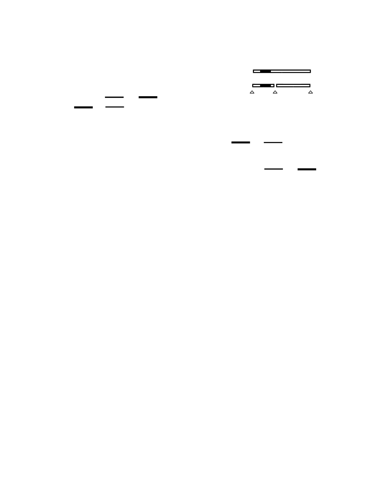
Figure 10.3
Schematic depiction of the molecular diagno-
sis of a genetic disease using an RFLP. The polymorphism
linked to the mutant gene has a novel endonuclease restric-
tion cleavage site. Southern blot hybridization shows a
shorter than normal DNA fragment in individuals with the
mutant gene. Triangles, restriction endonuclease cleavage
sites; black box, probe site.
NN
NM MM
Genotype
4
1.5
kb
Gene
N, normal
M, mutant
4 kb
1.5 kb
2.5 kb
Linked polymorphism


